Imaging Services
Our imaging services provide convenient, in-office CT scans, chest X-rays, and lung cancer screening to support the accurate diagnosis, early detection, and ongoing management of pulmonary conditions. By offering imaging directly within our practice, we are able to streamline care, reduce delays, and ensure imaging results are closely coordinated with your treatment plan. These services are available exclusively to our established patients, allowing for seamless communication and continuity of care within our clinic.
Types of Imaging Services
Chest X-rays and CT scans are important imaging tools used to evaluate the lungs, airways, and surrounding structures. Chest X-rays may include single-view or multi-view images and are commonly used to assess infections, fluid buildup, lung collapse, or changes over time. CT scans provide more detailed, cross-sectional images of the chest and may be performed with or without contrast to closely evaluate lung tissue, blood vessels, nodules, scarring, or other complex conditions. Together, these imaging services help diagnose pulmonary disease, guide treatment decisions, and monitor response to care with greater accuracy.
CT Scan
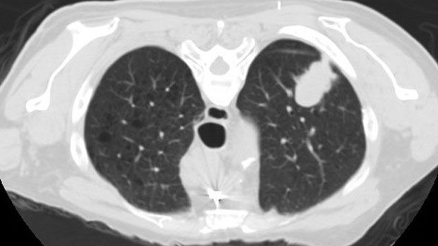
A CT scan (computed tomography) of the chest is performed to provide detailed, cross-sectional images of the lungs, airways, and surrounding structures that cannot be seen on a standard X-ray. It is commonly used to evaluate lung nodules, scarring, infections, blood clots, inflammation, and other complex pulmonary conditions, as well as to monitor known lung disease over time. During the scan, you will lie comfortably on a table that moves through the CT scanner while a series of images are taken; the test is painless and typically takes only a few minutes. In some cases, contrast dye may be used to enhance image clarity, and you will receive instructions beforehand if this is needed. After the scan, you can usually return to normal activities right away, and the images are reviewed to help guide diagnosis and treatment planning.
Chest X-Ray
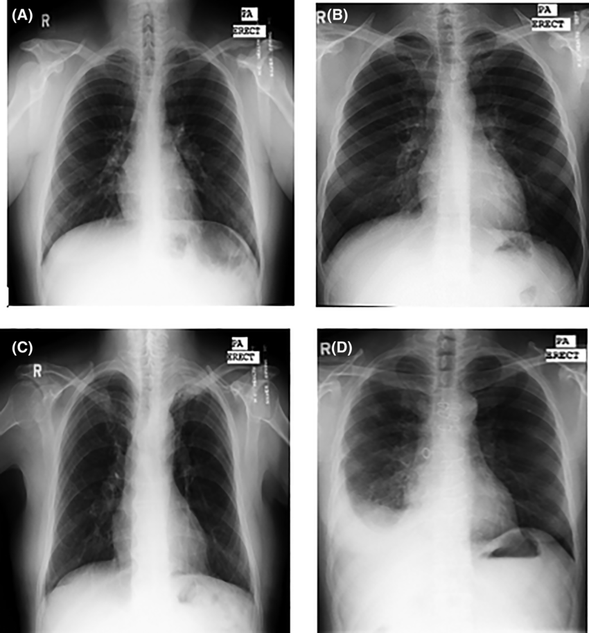
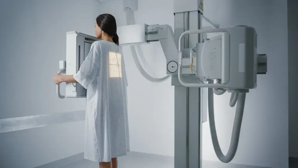
A chest X-ray is a quick, non-invasive imaging test used to evaluate the lungs, airways, heart, and surrounding structures. It is commonly performed to assess conditions such as infections, fluid around the lungs, lung collapse, chronic lung disease, or changes in symptoms over time. During the exam, you will be asked to stand or sit in front of the X-ray machine and briefly hold your breath while the images are taken. The test takes only a few minutes, involves minimal radiation exposure, and you can return to normal activities immediately afterward.
Lung Cancer Screening
Lung cancer screening is performed using a low-dose CT (LDCT) scan to detect lung cancer at an early stage, often before symptoms develop. The screening is recommended for individuals who meet specific risk criteria, such as a history of smoking, because early detection can significantly improve treatment options and outcomes. During the exam, you lie comfortably on a table that moves through the CT scanner while detailed images of the lungs are captured in just a few minutes. The test is painless, does not require contrast dye, and uses a lower amount of radiation than a standard CT scan. The images are carefully reviewed for lung nodules or other abnormalities, and results are used to determine whether routine annual screening, further imaging, or additional evaluation is needed.